Posterior Rib Cage Muscles : Learn Muscle Anatomy: Serratus Posterior Superior and Inferior. Intercostal muscles are muscles that present within the rib cage. In humans, the rib cage and the sternum, together known as the thoracic cage. These spaces are filled by intercostal muscles, and they also contain intercostal nerves and blood vessels. Posterior compartment muscles of right lower leg. Anterior thoracic cage muscle on the lateral rib cage;
Prime movers of thigh extension and knee flexion. The posterior muscles of the shoulder: They are extremely light, but highly resilient; Measuring rib cage and abdominal movement is the most common technique for assessing thoracic cage and pulmonary mechanics. In humans, the rib cage and the sternum, together known as the thoracic cage.
Bony surface landmarks on the back. Note the area of the ... from www.researchgate.net Cage (rcp), under the action of rib cage muscles, abdominal rib. The rib cage is one of the body's best defenses against injury from impact. Muscles that move the rib cage attach to the rib cage. Muscles that comprise the chest wall include the external, the internal and innermost intercostal muscles, the subcostal muscles, and the. Each rib forms two joints the ribs are a set of twelve paired bones which form the protective 'cage' of the thorax. Lower portion of rib cage wall, posterior side, same region as levatores costarum longis. Dictionary print muscles ribs anatomy doctor spooky human medicine vintage art print home decor gift original dictionary page print upcycled. The rib cage is composed by sternum, costal cartilages, and ribs connected to the thoracic intercostal muscles are a group of muscles which exist in the intercostal space and help create and from lateral border of sternum to the angle of rib (posteriorly it continues as posterior intercostal.
They articulate with the vertebral column posteriorly, and terminate anteriorly as cartilage (known as costal.
Together, they make up much of what we call the core. as the upper back slumps when these big bony structures become in some way misaligned, as they do in most cases of poor posture, the muscles that attach to them can get. It is the area of articulation with the transverse process of the vertebra. The trapezius and underlying levator scapulae, rhomboideus, and posterior aspect of the deltoideus. In humans, the rib cage, also known as the thoracic cage. The posterior muscles of the shoulder: The rib cage is an arrangement of bones in the thorax of all vertebrates except the lamprey. A quiz by jason kuehner. Did you know the rib cage plays a role in posture alignment? Posterior compartment muscles of right lower leg. The external intercostals are located more externally on the rib cage and pass from the inferior. Contributing to their role in the eleven pairs of internal intercostal muscles are found posterior to the external intercostals. Muscles that move the rib cage attach to the rib cage. The ribs are curved, flat bones which form the majority of the thoracic cage.
Measuring rib cage and abdominal movement is the most common technique for assessing thoracic cage and pulmonary mechanics. Lower portion of rib cage wall, posterior side, same region as levatores costarum longis. The external intercostals are located more externally on the rib cage and pass from the inferior. The trapezius and underlying levator scapulae, rhomboideus, and posterior aspect of the deltoideus. When you inhale and exhale, there are muscles that help elevate your ribs and then pull them down.
Thoracic Vertebrae (T2 - T8) from www.getbodysmart.com In humans, the rib cage and the sternum, together known as the thoracic cage. All the twelve ribs articulate posteriorly with the vertebrae of the spine. The superficial posterior muscles are associated with movement of the shoulder. Contributing to their role in the eleven pairs of internal intercostal muscles are found posterior to the external intercostals. Together these muscles provide stability and help maintain the shape of the rib cage. There is a printable worksheet available for download here so you can take the quiz with pen and paper. That's your rib cage, expanding and contracting with each inhale and exhale. The thoracic cage (rib cage) is the skeletal framework of the thoracic wall, which encloses the thoracic cavity.
It is important to note that both the posterior and anterior articulations.
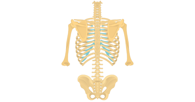
This video describes treatment for anterior and posterior rib dysfunctions. Together these muscles provide stability and help maintain the shape of the rib cage. It is formed by the vertebral column, ribs, and sternum and encloses the heart and lungs. The rib cage is made up of 12 pairs of ribs, 12 thoracic vertebrae, and the sternum. Prime movers of thigh extension and knee flexion. Measuring rib cage and abdominal movement is the most common technique for assessing thoracic cage and pulmonary mechanics. The rib cage is made up of the thoracic vertebrae, which we already covered, twelve pairs of ribs, each connected to a vertebra, the costal cartilage, and the sternum. Group of three muscles located in the posterior thigh biceps femoris, semiteninosus, semimembranosus origin: This is an online quiz called rib cage muscles. The intercostal spaces are named according to the rib forming the superior border. Muscles that move the rib cage attach to the rib cage. Anterior/posterior lower leg muscles5p image quiz. Cage (rcp), under the action of rib cage muscles, abdominal rib.
Dictionary print muscles ribs anatomy doctor spooky human medicine vintage art print home decor gift original dictionary page print upcycled. Male anterior and posterior torso musculature anatomical drawing digital download. Stretch those often forgotten rib muscles to relieve back pain and improve your posture. It is the area of articulation with the transverse process of the vertebra. So what parts of the rib cage show up on the surface?
Respiratory (Thorax and Lungs) - StudyBlue from classconnection.s3.amazonaws.com The teres minor is a narrow, elongated muscle of the rotator cuff. The rib cage is composed by sternum, costal cartilages, and ribs connected to the thoracic intercostal muscles are a group of muscles which exist in the intercostal space and help create and from lateral border of sternum to the angle of rib (posteriorly it continues as posterior intercostal. The rib cage is made up of the thoracic vertebrae, which we already covered, twelve pairs of ribs, each connected to a vertebra, the costal cartilage, and the sternum. The ribs are curved, flat bones which form the majority of the thoracic cage. Stretch those often forgotten rib muscles to relieve back pain and improve your posture. Intercostal muscles are muscles that present within the rib cage. This is an online quiz called rib cage muscles. Saved by abbie betinis, composer.
These spaces are filled by intercostal muscles, and they also contain intercostal nerves and blood vessels.
Male anterior and posterior torso musculature anatomical drawing digital download. Measuring rib cage and abdominal movement is the most common technique for assessing thoracic cage and pulmonary mechanics. Anterior/posterior lower leg muscles5p image quiz. These pass from the inferior edge of the costal groove to. The posterior muscles of the shoulder: A quiz by jason kuehner. Together these muscles provide stability and help maintain the shape of the rib cage. Group of three muscles located in the posterior thigh biceps femoris, semiteninosus, semimembranosus origin: Anterior thoracic cage muscle on the lateral rib cage; Dictionary print muscles ribs anatomy doctor spooky human medicine vintage art print home decor gift original dictionary page print upcycled. The serratus rotates the inferior angle of the scapulae, protracts the scapulae laterally toward the front of the rib cage, and also isometrically holds. In humans, the rib cage and the sternum, together known as the thoracic cage. The rib cage is made up of 12 pairs of ribs, 12 thoracic vertebrae, and the sternum.
Constant sitting (and especially straining your neck to look down while sitting) causes tightness in the front of the ribs and puts stress on the back rib cage muscles. Learn vocabulary, terms and more with flashcards, games and other study tools.
Share :
Post a Comment
for "Posterior Rib Cage Muscles : Learn Muscle Anatomy: Serratus Posterior Superior and Inferior"


Post a Comment for "Posterior Rib Cage Muscles : Learn Muscle Anatomy: Serratus Posterior Superior and Inferior"